Retinitis Pigmentosa (RP) is the name given to a group of inherited eye diseases that affect the light-sensitive rods and cones in the retina, It is a retinal degenerative disease characterized by pigment deposits predominant in the peripheral retina and by a relative sparing of the central retina. In most of the cases of RP, there is a primary degeneration of the photoreceptor rods, with secondary degeneration of cones (Beshare, 2011). Thus, the typical RP is also described as a rod-cone dystrophy, photoreceptor rods being more affected than cones. This sequence of photoreceptor involvement explains why patients initially present with night blindness, and only in the later life would suffer visual impairment in diurnal conditions. Retinitis pigmentosa (RP) was first described and named by Donders in 1857(Donders,1857). This diagnosis now includes a heterogeneous group of inherited retinal diseases,the precise number of which vary according to the inclusion criteria defined by different investigators (Heckenlively 1982). RP is considered to be one of the most frequent causes of blindness during working life in the industrialized countries.

Classification
One often confounding feature of RP is the many different ways it can be classified. RP can be classified by its mode of inheritance, age of onset, fundus appearance, pattern of functional vision loss or by genetic mutation.
• Most common genes causing retinitis pigmentosa include USH2A,PDE6B,PDEGA,MYO7A,CRB1,RGR(Autosomal recessive),Rho,RP1,RDS,ROM(Autosomal dominant),RPGR,RP2(X-linked RP).
• By Age Of Onset- Severe forms of RP manifest before the first year of life referred to as Leber Congenital Amaurosis(LCAA) . The forms of the RP occurring between 1 and 5 years have been termed as Juvenile RP or Severe early childhood-onset retinal dystrophy((SECORD).
• By Fundus appearance- Deviations from this classic fundus appearance have given rise to several alternative terms for pigmentary retinopathies including inverse RP, concentric RP, sector RP, retinitis punctate albescens, fundus albipunctatus, RP with preserved peri-artetriolar RPE, pigmented perivenous retinochoroidal atrophy and retinitis sine pigmento.
• By Functional loss-Historically, adult RP was categorized as either Type 1 (rod dysfunction) or Type 2 (rod and cone dysfunction) based on psychophysical testing .These classifications were further refined on the basis of electrophysiological findings to include the categories; rod-cone dystrophy, cone-rod dystrophy or cone dystrophy. Most forms of RP are Rod-cone dystrophies in which rod photoreceptor death occurs first and is later followed by subsequent cone photoreceptor death.
Signs/Symptoms
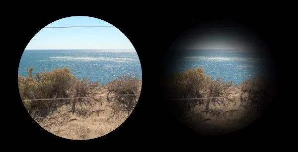
Retinitis Pigmentosa usually starts in childhood or adolescence. But exactly when it starts and how quickly it gets worse varies from person to person. Most people with RP lose much of their sight by early adulthood. Then by age 40, they are often legally blind. Because rods are usually affected first, the first symptom you may notice is that it takes longer to adjust to darkness (night blindness).In children, parents may comment that their child is afraid or becomes distressed in the dark. Older children often comment on poor vision compared with their fully sighted peers. In adults , difficulties with night driving are frequent. The second cardinal symptom of RP is progressive peripheral visual-field loss. Since central vision is spared early in the course of the disease, some patients do not notice this loss of visual field until the degeneration has become quite advanced. In later stages, your cones may be affected. That will make it harder for you to do detail work, and you may have trouble seeing colors. It’s rare, but sometimes the cones die first. Because there are many gene mutations that cause the disorder, its progression can differ greatly from person to person. You might find bright lights uncomfortable, a symptom called Photophobia. You also may start to see flashes of light that shimmer or blink. This is called Photopsia. Patients with cone and cone-rod dystrophies can present with early photophobia, decreased central vision and impaired color vision. These patients will typically have worse visual acuity than patients with rod-cone dystrophies due to earlier involvement of macular cones. Refractive errors in patients with RP have been studied and on average these patients are more myopic and have a greater degree of astigmatism than those in the general population.
Treatment
Currently, there is no known cure for most forms of RP ,although some treatments have been shown to slow down the progression of the disease and future therapies are promising. It is advisable to see you optometrist as soon as possible once any of these symptoms are notice.

What is @bettervision about?
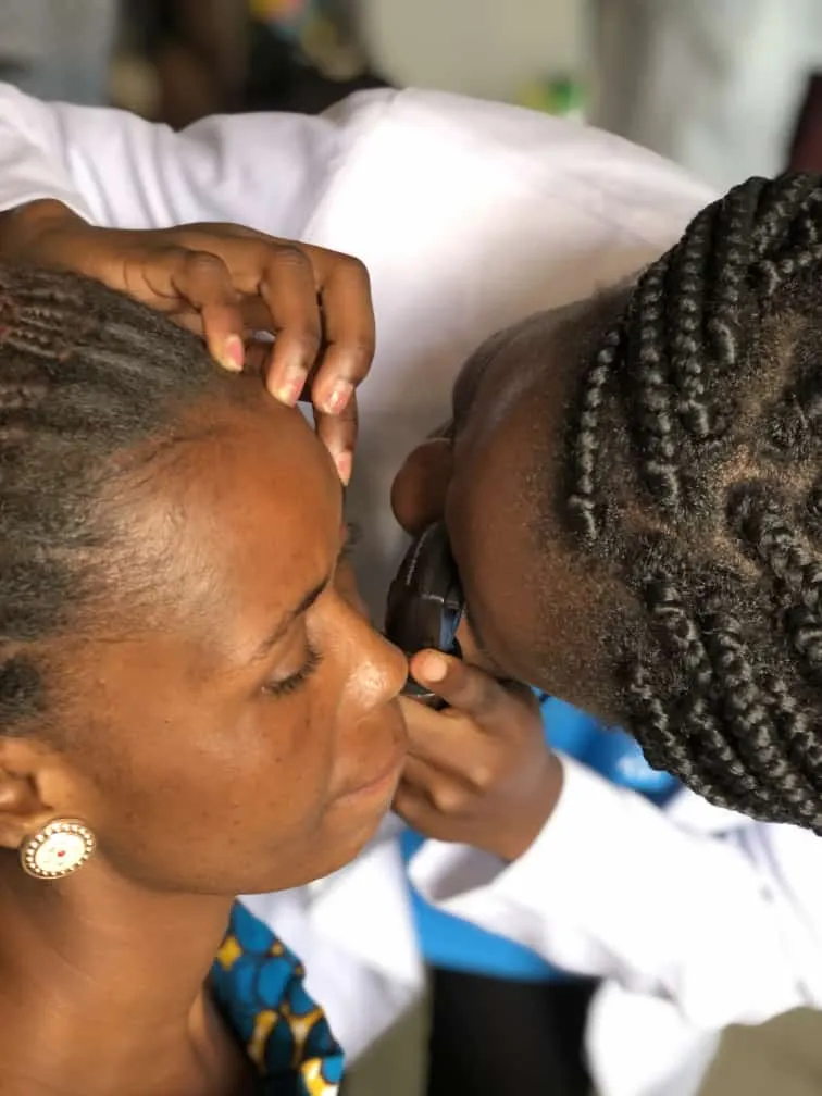
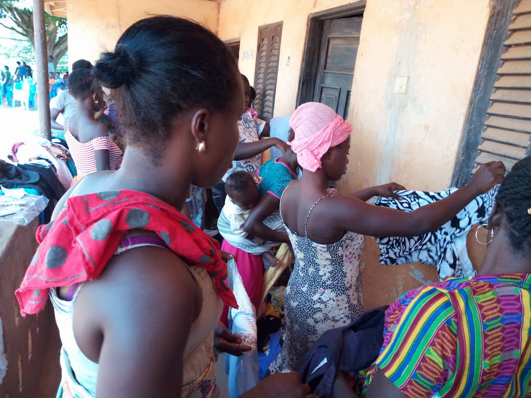
@bettervision is is a project initiated by @nattybongo and friends to give back to the society the knowledge and skill acquired through the Optometric Studies in Kwame Nkrumah University of Science and Technology, Ghana.It is an outreach system where we visit the less privileged communities to offer free eye screening services and education to the people within the community
AIMS AND OBJECTIVES
To reduce or prevent vision loss through diseases such as glaucoma, cataract and refractive errors.
To enlighten the majority of the Ghanaian population about the importance of proper visual care.
To conscientize people on the need for regular eye checks
To get more people to have their wards screened within the Critical periods of a Child’s Vision Development; thus from ages 3 to till about 10 years.
To help the blind and people with low vision live a better life within the society through education of the general public to stop stigmatization.
To help in the fight of extreme poverty that puts the health of people at risk
Our greatest gratitude goes to @fundition @adollaraday @surfyogi @girlsfoundation @bleepcoin @ackza @indigoocean @nanzo-scoop @steemstem @demotruk @pennsif @steem-ambassador @kasho and @wafrica for helping to make the aims and objectives of @bettervision a reality.


shirt.jpg](https://images.ecency.com/p/3jpR3paJ37V8JxyWvtbhvcm5k3roJwHBR4WTALx7XaoRovbdkGwm8sDfo9UUXEuNZXBMC1kdmJ6coMxZnzE8tqnHbG5dNEHKxuT43r9nisaaZ4NderHKyDtCXn8wp1tG6F6zi.png?format=match&mode=fit)