Hello everyone, how are you all? i hope you all are doing well and but i am not feeling well and my heart is not happy at all. its ok today we are continuing the endoscopy and today i would like to show endoscopic instruments and its uses and the procedure and parient was prepared. So i am going to share images and will explain.
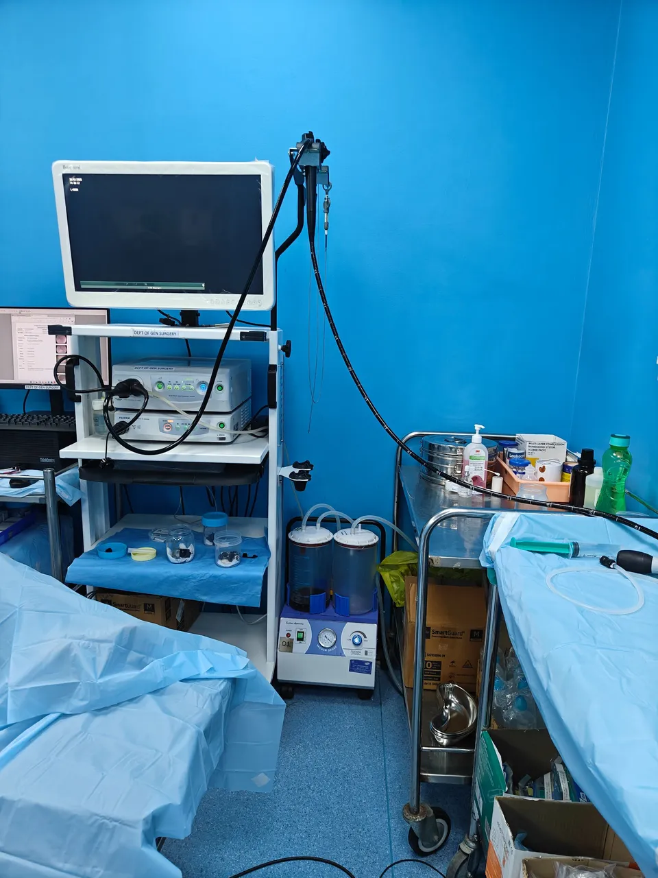
Endoscopy trolley
Endoscopic trolley is having video monitor, light source, video processor and
endoscope hanger.

First we will make the patient ready that patient should follow 6-8 hrs of fasting before endoscopy procedure. Then consent is taken from the patient or their relatives and next patient was asked to open their mouth this 10% lignocaine spray was given two puffs were given to reduce pain while inserting endoscopy.
10% Lignocaine spray
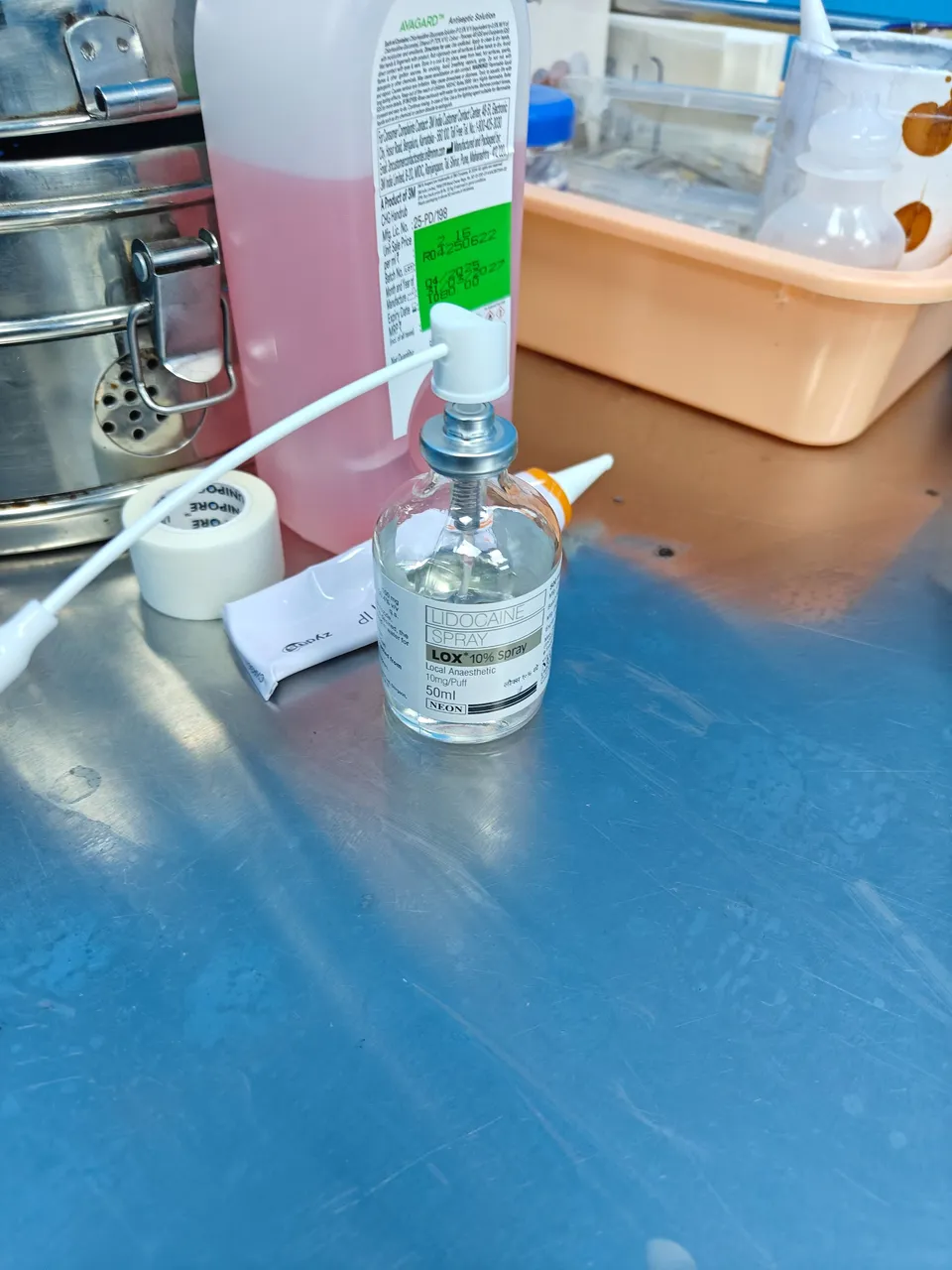
The patient should swallow it show that patient won't find any difficult while doing procedure.
Mouth Guard
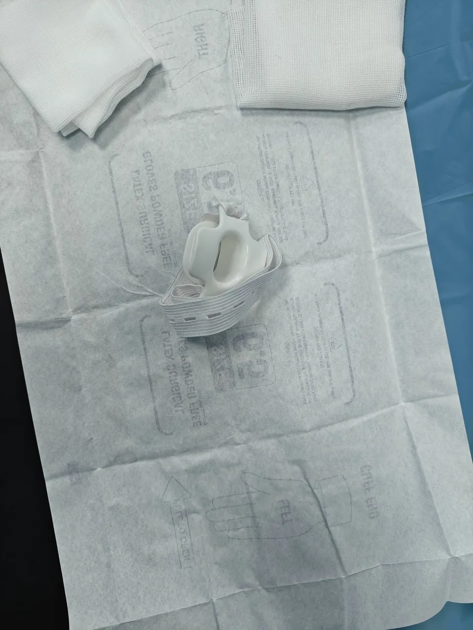
The patient is then asked to hold the with their teeth and this one is for insertion of endoscopy and to protect the endoscopy from biting by patients. Then patient asked to lie down in left lateral position
Esophagogastroduodenoscope
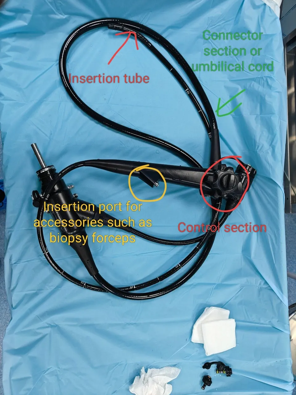

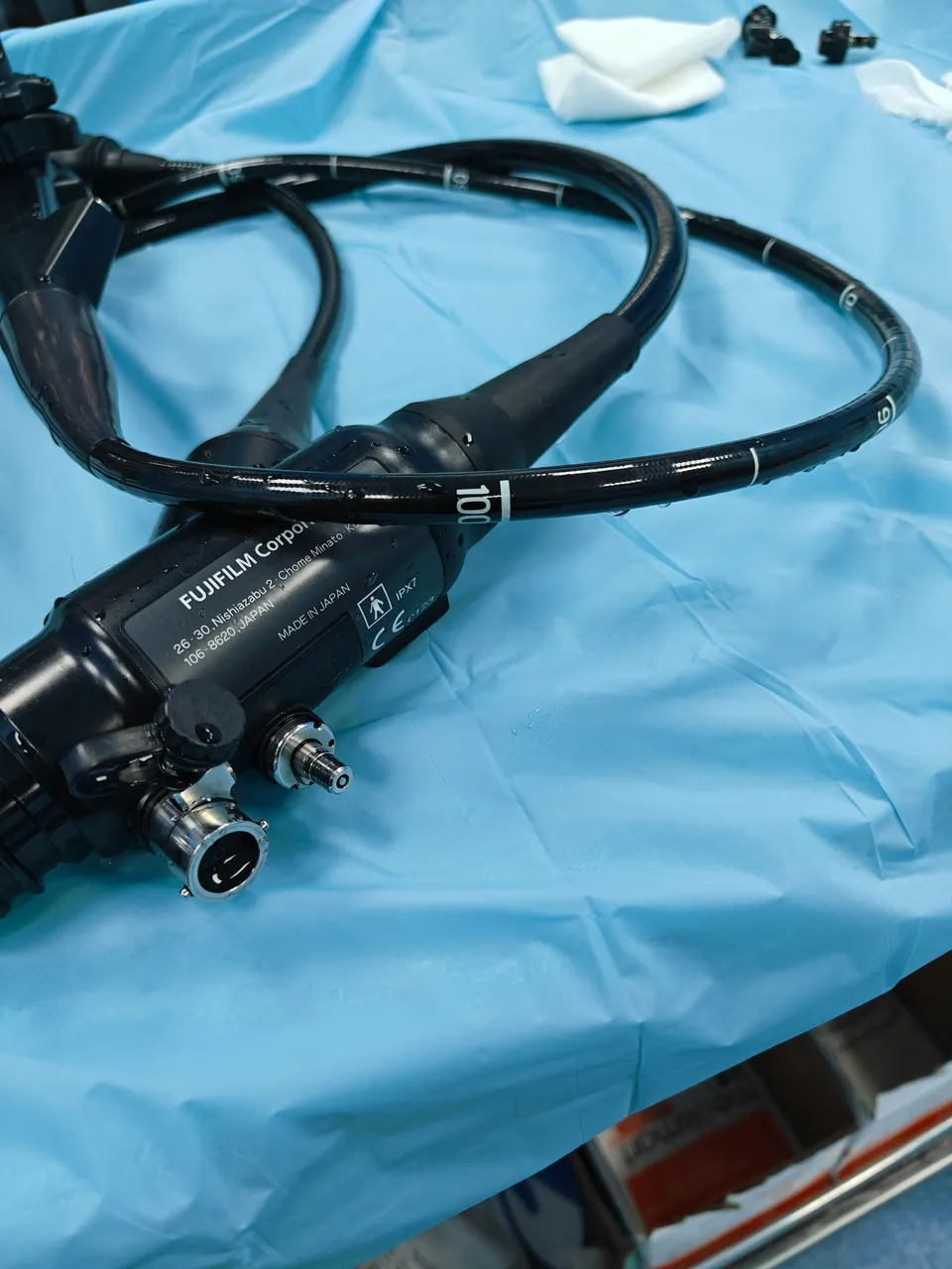
The above one is called Esophagogastroduodenoscope and it is also called upper GI endoscopy instrument and you can see it has biopsy port and its length about 100-110 cm and pediatrics will be 90 to 110 cm and it will be measured from upper incisor teeth. The length of the gastroesophageal junction is measured from upper incisor teeth and any abnormalities like barrets esophagus, candidiasis and malignancy is seen in the esophagus first. So basically we will examine the esophagus and gastroesophageal junction from incisor teeth. Then endoscopy is entered into stomach and the the mucosa of stomach is examined, any benign malignant lesions are there. In stomach, first we will see body of stomach then will go for antrum, pylorus and then we will enter into duodenum through sphincter and here we can check whether the scope is entering into duodenum or not. If its not entering then gastric outlet obstruction can be considered. Before entering into duodenum we will check antrum and body of stomach and most of the biopsies are taken from this area. If we are suspecting H pylori infection biopsy taken from antrum, pylorus or body and 1-2 biopsies will be take for RUT test. Ok now we entered into duodenum then in duodenum well check any growth or lesions are there and we will check ampulla of vater where common bile
Duct and pancreatic ducts opens in to duodenum. The endoscopy will reach to 2nd part of duodenum or 3rd part of duodenum in very rare cases. Beyond that we will go for enteroscopy or video capsule endoscopy.
Cytology brush
You can see feathery type bristles. This is used for cytological examination.

Endoscopy biopsy forceps
This is used to take biopsy.

I hope, you understood very well about endoscopy instruments and its uses and procedure. Still many things are there learn so be enthusiastic and awaited.
References
- Surgical Pathology of the Gastrointestinal System,Volume I,Prasenjit Das · Kaushik Majumdar Siddhartha Datta Gupta
Thanks for reading,
With regards,