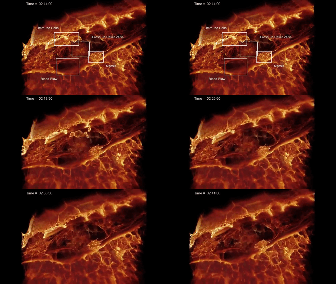
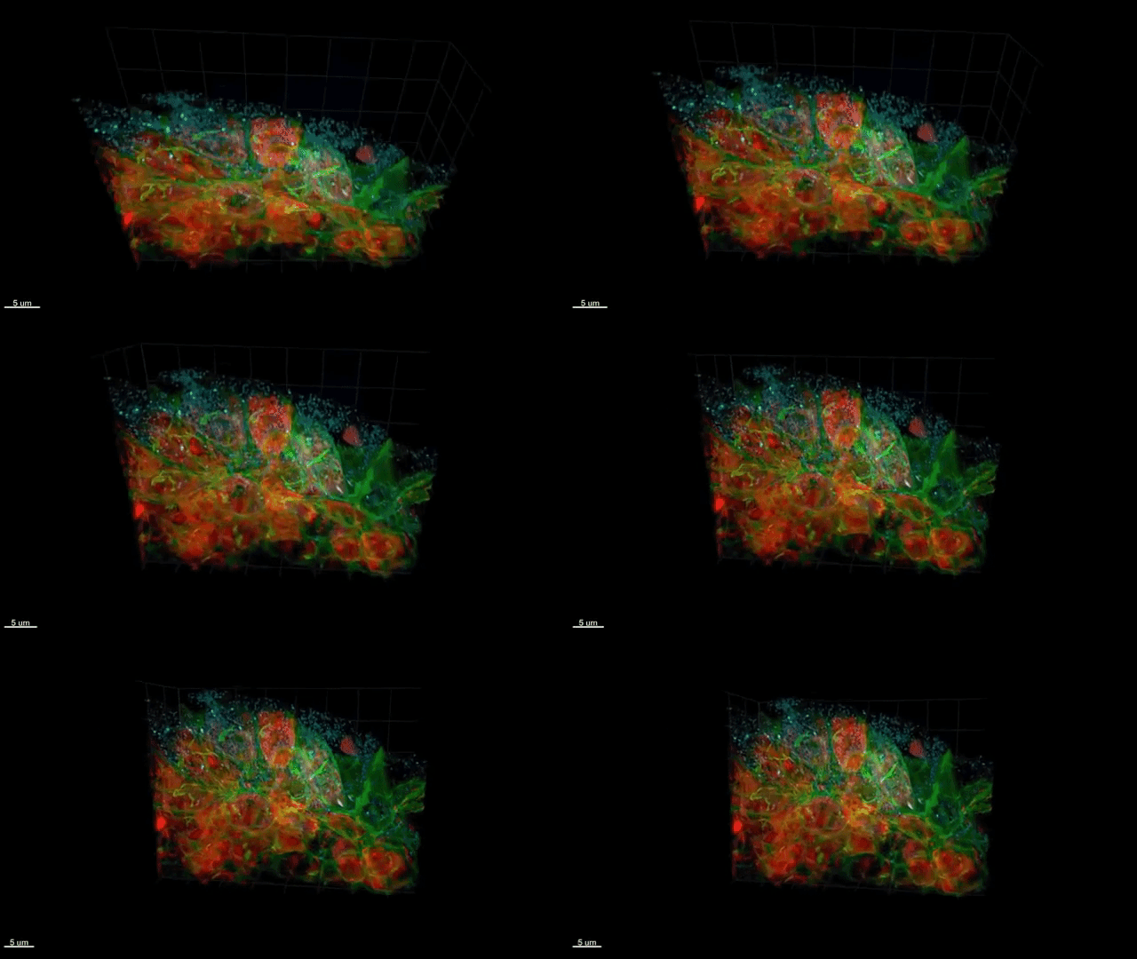
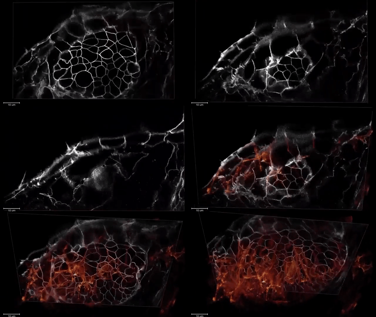
This is an incredible microscope, which has captures 3D images of sub-cellular movement into multi-cell organisms.

The HHMI Howard Hughes Medical Institute Leader Eric Betzig and his team of researchers have created this amazing microscope.
They have caught the high resolutions of dynamic 3D images and footage of the movement.

They can see the sub-cellular movement deep within the multi-cellular organisms.
This team is able to see that it could never be seen before. It is a combination of features like 'Sheer lattice Microscopy' with 'Adaptive Optics'.

It is a remarkable testimony of how immune cells develop within the zebra fish embryo.
This example of this footage shows Zebra Fish in the immune cell migration in the inner ear.
image-video-sources-https://laughingsquid.com/3d-images-of-subcellular-movement - https://youtu.be

