
About twenty years ago I showed my doctor a spot behind my left ear. The doctor referred me to a dermatologist who performed a biopsy. The report came back: pemphigus vulgaris. I looked that up on the Internet and refused to accept the idea, so I went to a research hospital. Second biopsy: pemphigus vulgaris.
A powerful topical steroid erased the spot quickly and it never came back. As you read this blog you will see that my fortune was good in the extreme. Pemphigus can run a cruel course, and it can take a life. Many, many people who receive the same pathology report I did, do not share my good fortune.
Here's some of what I learned about pemphigus in the ensuing years. The information might be useful someday, for you, your family or your pet. Pemphigus affects animals, too.

Certain dog breeds are more susceptible to pemphigus than others. These breeds include Akita, Chow Chow, and Cocker Spaniels.
What is It?
According to Merk Manual:
"Pemphigus vulgaris is an uncommon, potentially fatal, autoimmune
disorder characterized by intraepidermal blisters and extensive erosions
on apparently healthy skin and mucous membranes". 1
Translated into ordinary language: pemphigus is very rare, and it can be fatal. Blisters form within the cells of the upper layer of skin. With a particular type of pemphigus, pemphigus vulgaris, blisters also form in the mucous membranes.
Pemphigus is categorized as an orphan disease by the U.S. Food and Drug Administration. The occurrence of pemphigus varies by region across the globe, and also by ethnicity. It ranges between 0.7 and 5 people, per million. Populations with a Mediterranean or Ashkenazi Jewish background are at higher risk. The incidence in these populations may be as high as 6-32 cases per million. A particular subset of pemphigus (endemic pemphigus) occurs with greater frequency in the Brazilian rainforest and in Colombia.
There are photos available on the Internet, of people afflicted with pemphigus. I'd feel I was violating those people if I used such distressing, personal photos. However I did find an illustration that is less intrusive and yet reflects how extensively pemphigus can affect the skin. This picture shows a patient (in 1896) with pemphigus foliaceus, PF, a less fatal type of the disease than pemphigus vulgaris, PV.
The severe condition of this patient is one that was suffered by most before the introduction of modern treatments.

Types of Pemphigus
As I poured through the literature, the words pemphigus and pemphigoid appeared in different contexts. A variety of blistering diseases can be characterized by the words pemphigus or pemphigoid. I found some categorical clarity in a 2016 article, Mechanisms of Disease: Pemphigus and Bullous Pemphigoid, published in the Annual Review of Pathology. The authors separate the histological character (tissue analysis, available from a biopsy) of the disease from its clinical manifestations (what does it look like, what are the symptoms).
According to this article (and also according to the International Pemphigus and Pemphigoid Foundation), histologically there are two types: pemphigus vulgaris--PV-- and pemphigus foliaceus--PF . In tissue samples of both types there will be evidence of autoantibodies.. It is the type of autoantibody and its specific target tissue that become diagnostic in pemphigus. A pathologist assesses this in a biopsy.
In pemphigus, autoantibodies target a specific substance found in the epidermis: desmoglein. With the destruction of desmoglein, essential cells, called keratinocytes, cannot hold together. Blisters form in those areas of failure and the skin essentially falls apart.
Keratinocytes

Keratinocytes make up about 95% of the epidermis. Without treatment, a pemphigus patient may actually lose most of the epidermis. This would lead to fluid loss, widespread infection, and likely sepsis. Most patients would die in about two years.
There are different types of desmoglein. A diagnosis of PV or PF is based on which kind of desmoglein is under attack. A biopsy will reveal this. If it turns out that desmoglein 1 is the target, then the finding is for PF. If desmoglein 3 is under attack, then the diagnosis is PV. There are other diagnostic tests that are used to come up with a definitive diagnosis, but separating the histology into attacks on desmoglein 1 and 3 is a basic step.
IgG, an Autoantibody Found in Pemphigus

The presence of IgG autoantibodies is a hallmark of PF and PV.
IgG is an immunoglobulin. It is the most abundant immunoglobulin in the body and is necessary for proper function of the immune system. As a matter of fact, it is the only antibody class that can cross the placental barrier. This allows the mother to bestow some of her immunity on the fetus.
IgG is not the only autoantibody involved in the development of pemphigus, but it is the most predominant. It is also implicated in a number of other autoimmune diseases.
Desmoglein 1 and Desmoglein 3
Most of us know that keratin is contained in hair, nails and skin. Keratinocytes manufacture and store keratin, and are the basic building blocks of the epidermis. Keratinocytes are held together by extracellular structures called desmosomes. These structures are like the mortar between bricks in a wall. Desmosomes are comprised of two glycoproteins, desmoglein and desmocollin.
Remember, in PF and PV, it is desmoglein that is destroyed by autoantibodies. So, if the desmoglein is destroyed, desmosome, the intracellular mortar between keratinocytesis is destroyed. The keratinocytes are no longer held together. The basic building blocks of the skin separate, and the skin literally falls apart. Blisters form in the gaps that open up.
I made a GIF to demonstrate how the structure of the skin is undermined by the destruction of desmoglein. Here the mortar between the bricks in a wall slowly erodes and the bricks fall away. Compare this GIF to the image of tissue undergoing the process of acantholysis, a little further down in the blog.
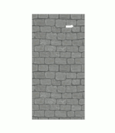
Illustration of Desmosome Cell Adhesion

Desmosomes are only one type of intracellular juncture in animal tissue. Most of us are more familiar with another kind: cartilage. Both desmosomes and cartilage are designed to bear weight and to allow for flexibility. You can see in the illustration above that fibers connect one cell to another at an adhesion point called a cadherin. It is the cadherin that is is made up of desmoglein. This is precisely the area of vulnerability in pemphigus.
Below is a microscopic image of tissue taken from a patient who has PV. Even with the untrained eye, we can see gaps in the tissue. According to the description that appears under this photo, "an early intraepidermal vesicle" is evident in this tissue.2 That is, blisters are already forming, and are evident. Compare this gap to the gap in my brick wall GIF.
The process by which desmosomes are destroyed and epithelial tissue separates is called acantholysis.

A Personal Digression
In the title of this blog I refer to a personal perspective. Here it is: my experience does not resemble in any way what I have just described. Despite the diagnosis of pemphigus vulgaris, based on two biopsies, a blister never formed. Both pathologists did find that IgG autoantibodies were attacking desmoglein 3 in the biopsied tissue. By the time I showed my doctor the spot, almost two years had passed (!).
The spot had grown a bit, but it was still under 2 cm. I thought I might have a modest squamous cell carcinoma.
I was wrong.

I selected this diagram out of many because it indicates that squamous cells are near the surface of the skin. Stratified squamous epithelium comprise the epidermis. It is here, in the squamous cells, that pemphigus (PV and PF) develops. These cells are flat (wider than they are high) and are arranged in layers.
The image below shows the lining of a normal esophagus. Stratified squamous cells are visible. The esophagus is lined with a mucous membrane, which means it is vulnerable to the development of PV, but not PF.

In PV, mucous membranes are often the first sites affected. Typically, oral lesions appear. Because of this, dentists can play an important role in detecting the disease. Mucosal involvement may extend to the esophagus, the nose and even inside the eyelids. (Both pemphigus vulgaris and pemphigus foliaceus may effect the eyelids.)
Before the advent of modern treatment, oral and esophageal lesions inhibited eating, and this contributed to mortality.
Clinical Manifestations of Pemphigus
While pemphigus is broadly described as being either pemphigus foliaceus or pemphigus vulgaris, based on histology, there are subdivisions of these two types. It's hard to get different sources to agree on what these subdivisions are, so I settled for the grouping offered by NORD, National Organization for Rare Diseases. Please note, these subdivisions may overlap, or appear sequentially.
NORD lists six subdivisions, two of these have already been discussed in some detail:
Pemphigus Foliaceus
Pemphigus Vulgaris
I'll say a few words about the other four subcategories:
Endemic Pemphigus
I referred to this earlier in the blog as occurring in the rainforests of Brazil and Colombia. I have also read an article which relates similar cases in Peru to the same type of endemic disease. The suspicion has always existed that an environmental factor triggers the disease in genetically susceptible individuals. A fascinating 2016 article in the Journal of Immunology suggests that the environmental exposure might be the bite of a sandfly. In this case, pathogenic IgG4, instead of IgG1 (which is often the culprit) is implicated.

Sandflies are found all over the world, but this variety is only found in the so-called 'New World'--which would encompass Brazil, Peru and Colombia, the countries where endemic pemphigus has been identified.
Pemphigus Erythematosus (Senear-Usher syndrome)
This may be an overlap disease with lupus erythematosus. Patients have serology that is positive for lupus (ex: anti-SM and double-stranded DNA antigens, hallmarks of lupus) and also have histology and clinical presentation compatible with pemphigus.
Pemphigus Vegetans
This is described by most sources as a rare, localized, benign form of PV. Partly because of its rarity, it can be difficult to diagnose.
Drug-Induced Pemphigus

The -SH in the formula above is a sulfhydryl, and it is this that characterizes the thiol group of drugs. This group of drugs is implicated in the development of pemphigus.
This is probably one of the most important parts of my blog today: pemphigus may be caused by drugs that are commonly prescribed. In many cases, the disease will clear up when the drug is withdrawn, but this is not always the case. Also, pemphigus may not develop until months after the drug has been commenced, so it's hard to draw a relationship between the drug and the disease. According to StatPearls, drugs are actually the most likely cause of pemphigus, especially pemphigus foliaceus.
Certain classes of drugs are more closely associated with the disease than others. Thiols (especially d-penicillamine) have been implicated. Phenols have also been implicated. Interestingly, diet can also be a source of phenols and thiols, so foods that contain these...chives, garlic, onion, black pepper, cashew and mangoes...should be avoided by anyone who has been diagnosed with pemphigus.
According to DermNet NZ, about 50% of the cases caused by thiols will clear up when the drug is withdrawn. Disease caused by non-thiol drugs, "have a better prognosis".3
Anti-hypertensives and NSAIDs have also been implicated.
Treatments
The cornerstone of treatment remains corticosteroids, administered systemically, topically or a combination of the two. These drugs can have serious side effects, including: cataracts, diabetes, osteoporosis, psychosis, chronic migraine and increased susceptibility to infection. The effects have been found to be dose dependent. The goal, for physician and patient, is to take the lowest effective dose. This treatment strategy is referred to as 'steroid sparing', and it often involves a balancing act between drug efficacy and drug effects.
Some other treatments for pemphigus are:
Azathioprine
Cyclophosphamide
Mycophenalate
Methotrexate
Rituximab
Dapsone
IV infusion of immunoglobulin
Plasmapheresis
Causes/Antagonists
We've already covered drugs and diet. Genetics seems to play a weak role, particularly in some families. According to GARD, Genetic and Rare Diseases Information Center, " Predisposition...is linked to genetic factors...histocompatibility complex (MHC) class II molecules, in particular alleles of human leukocyte antigen (HLA) DR4, appear to increase susceptibility to pemphigus vulgaris."
There are known environmental antagonists, besides drugs, diet and, possibly, sandflies. One of these, very avoidable, is the sun..or any form of UV radiation.
Ending on a Personal Note
I have a serious UV allergy, which encompasses not only sun but indoor sources of UV. This may be one of the factors that has kept pemphigus at bay all these years. I avoid the sun. My wardrobe offers evidence of that. I have a closet full of hats. So I decided to end the blog on a rather humorous note: a GIF that shows just some of my hats :)

Anyone who wants to use any of my GIFs, for any reason, may do so without restriction.

Footnotes
1Merck Manual Professional Version: Pemphigus Vulgaris
3DermNet Drug-induced pemphigus
Some Sources Used in Writing This Blog
Sequence Characterization of DSG3 Gene to Know Its Role in High-Altitude Hypoxia Adaptation in the Chinese Cashmere Goat https://www.frontiersin.org/articles/10.3389/fgene.2018.00553/full
Companion Animals, Too, Can Get Pemphigus http://www.pemphigus.org/companion-animals-too-can-get-pemphigus-2/
Pemphigus in Dogs https://vcahospitals.com/know-your-pet/pemphigus-in-dogs
Pemphigus Vulgaris https://www.merckmanuals.com/professional/dermatologic-disorders/bullous-diseases/pemphigus-vulgaris
Orphan Products: Hope for People With Rare Diseases https://www.fda.gov/drugs/drug-information-consumers/orphan-products-hope-people-rare-diseases
Pemphigus http://www.pemphigus.org/research/clinically-speaking/pemphigus/
Mechanisms of Disease: Pemphigus and Bullous Pemphigoid https://www.ncbi.nlm.nih.gov/pmc/articles/PMC5560122/
Nature and functions of autoantibodies https://www.ncbi.nlm.nih.gov/pmc/articles/PMC2703183/
Layers of the Skin http://library.open.oregonstate.edu/aandp/chapter/5-1-layers-of-the-skin/
Usefulness of desmoglein 1 and 3 in serodiagnosis of pemphigus vulgaris and its correlation with disease activity - ELISA study http://www.jofs.in/article.asp?issn=0975-8844;year=2014;volume=6;issue=2;spage=104;epage=107;aulast=Gandhi
Definition of human autoimmunity--autoantibodies versus autoimmune disease https://www.ncbi.nlm.nih.gov/pubmed/19963079
Immunoglobulin G https://www.sciencedirect.com/topics/neuroscience/immunoglobulin-g
Natural Antibodies Bridge Innate and Adaptive Immunity http://www.jimmunol.org/content/jimmunol/194/1/13.full.pdf
Serum IgG Subclasses in Autoimmune Diseases https://www.ncbi.nlm.nih.gov/pmc/articles/PMC4602543/
Kinetin Improves Barrier Function of the Skin by Modulating Keratinocyte Differentiation Markers
https://www.ncbi.nlm.nih.gov/pubmed/28223740Desmocollin and desmoglein – different roles in desmosomes http://jcs.biologists.org/content/127/10/e1001
Learn more about Acantholysis https://www.sciencedirect.com/topics/medicine-and-dentistry/acantholysis
Potential Risk Factors for Cutaneous Squamous Cell Carcinoma include Oral Contraceptives: Results of a Nested Case-Control Study https://www.ncbi.nlm.nih.gov/pmc/articles/PMC2872290/
Pemphigus vulgaris presenting as gingival involvement https://www.ncbi.nlm.nih.gov/pmc/articles/PMC3505431/
When the Eyes Have It http://www.pemphigus.org/when-the-eyes-have-it-2/
Endemic pemphigus in the peruvian Amazon: epidemiology and risk factors for the development of complications during treatment http://www.scielo.br/scielo.php?script=sci_arttext&pid=S0365-05962012000600003
Overlapping IgG4 Responses to Self- and Environmental Antigens in Endemic Pemphigus Foliaceus http://www.jimmunol.org/content/196/5/2041
A New Classification System for IgG4 Autoantibodies https://www.ncbi.nlm.nih.gov/pmc/articles/PMC5816565/
Autoantibodies in Senear-Usher Syndrome: Cross-Reactivity or Multiple Autoimmunity?
https://www.ncbi.nlm.nih.gov/pmc/articles/PMC3539423/Pemphigus vegetans: An unusual presentation https://www.ncbi.nlm.nih.gov/pmc/articles/PMC3505428/
Drug Induced Pemphigus https://www.ncbi.nlm.nih.gov/books/NBK499864/
Drug-induced pemphigus https://www.dermnetnz.org/topics/drug-induced-pemphigus/
Antihypertensives in dermatology Part II - Cutaneous adverse reactions to antihypertensives http://www.ijdvl.com/article.asp?issn=0378-6323;year=2018;volume=84;issue=2;spage=137;epage=147;aulast=Ranugha
Pemphigus vulgaris https://rarediseases.info.nih.gov/diseases/7355/pemphigus-vulgaris
Adverse effects of oral corticosteroids in relation to dose in patients with lung disease https://www.ncbi.nlm.nih.gov/pmc/articles/PMC1746020/
Patterns of steroid and steroid sparing regimens among older inflammatory bowel disease (IBD) patients with contraindications to tumor necrosis factor antagonists (ANTI-TNFS) https://www.valueinhealthjournal.com/article/S1098-3015(14)00294-0/fulltext
Azathioprine in the treatment of Autoimmune Blistering diseases http://www.pemphigus.org/azathioprine-in-the-treatment-of-autoimmune-blistering-diseases/
A comparative effectiveness research of azathioprine and cyclophosphamide on the clinical and serological response in pemphigus vulgaris http://www.e-ijd.org/article.asp?issn=0019-5154;year=2016;volume=61;issue=4;spage=418;epage=426;aulast=Sardana
Medline ® Abstract for Reference 5 of 'Initial management of pemphigus vulgaris and pemphigus foliaceus' https://www.uptodate.com/contents/initial-management-of-pemphigus-vulgaris-and-pemphigus-foliaceus/abstract/5
Methotrexate in the treatment of pemphigus vulgaris: experience in 23 patients https://www.ncbi.nlm.nih.gov/pubmed/23772610
FDA Approves Genentech’s Rituxan (Rituximab) for Pemphigus Vulgaris
https://www.gene.com/media/press-releases/14727/2018-06-07/fda-approves-genentechs-rituxan-rituximaDapsone in the treatment of pemphigus vulgaris: adverse effects and its importance as a corticosteroid sparing agent https://www.ncbi.nlm.nih.gov/pmc/articles/PMC4540507/
Effect of Intravenous Immunoglobulin Therapy on Serum Levels of IgG1 and IgG4 Antidesmoglein 1 and Antidesmoglein 3 Antibodies in Pemphigus Vulgaris https://jamanetwork.com/journals/jamadermatology/article-abstract/420177
Plasmapheresis in Refractory Pemphigus Vulgaris: Revisiting an Old Treatment Modality Used in Synchrony With Pulse Cyclophosphamide https://www.mdedge.com/dermatology/article/97747/wounds/plasmapheresis-refractory-pemphigus-vulgaris-revisiting-old
Pemphigus vulgaris https://rarediseases.info.nih.gov/diseases/7355/pemphigus-vulgaris
Sun Exposure http://www.pemphigus.org/tag/sun-exposure/
Photosensitivity https://www.ncbi.nlm.nih.gov/books/NBK431072/